
VR Module · Anatomy · Healthcare
Human Heart Anatomy
Cardiovascular System
See it
Inside the module.
Learning objectives
By the end of this module, students will be able to:
Identify the major parts of the heart - atria, ventricles, valves, septum, aorta, vena cava, pulmonary artery, and pulmonary veins
Understand the internal structure of the heart through sectional and inside views
Explain the path of oxygenated and deoxygenated blood through the heart
Understand the role of valves in maintaining one-way blood flow
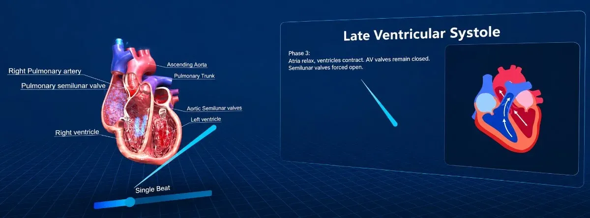
Visualise the basic cardiac cycle - contraction, relaxation, filling, and pumping
Relate heart anatomy to physiology, circulation, and common clinical concepts
The whole heart, in your hands.
The human heart is a four-chambered muscular pump at the centre of the cardiovascular system, and its three-dimensional structure is hard to grasp from flat diagrams. This module presents the heart as a complete, manipulable 3D model that students can rotate, section, and step inside.
Learners move between external anatomy, sectional inside views, and isolated structures - following oxygenated and deoxygenated blood through the chambers and great vessels, and seeing how the valves keep flow moving in one direction.
Why a 3D heart rather than a diagram?
- The heart is a three-dimensional pump, and a flat diagram forces students to reconstruct it mentally every time.
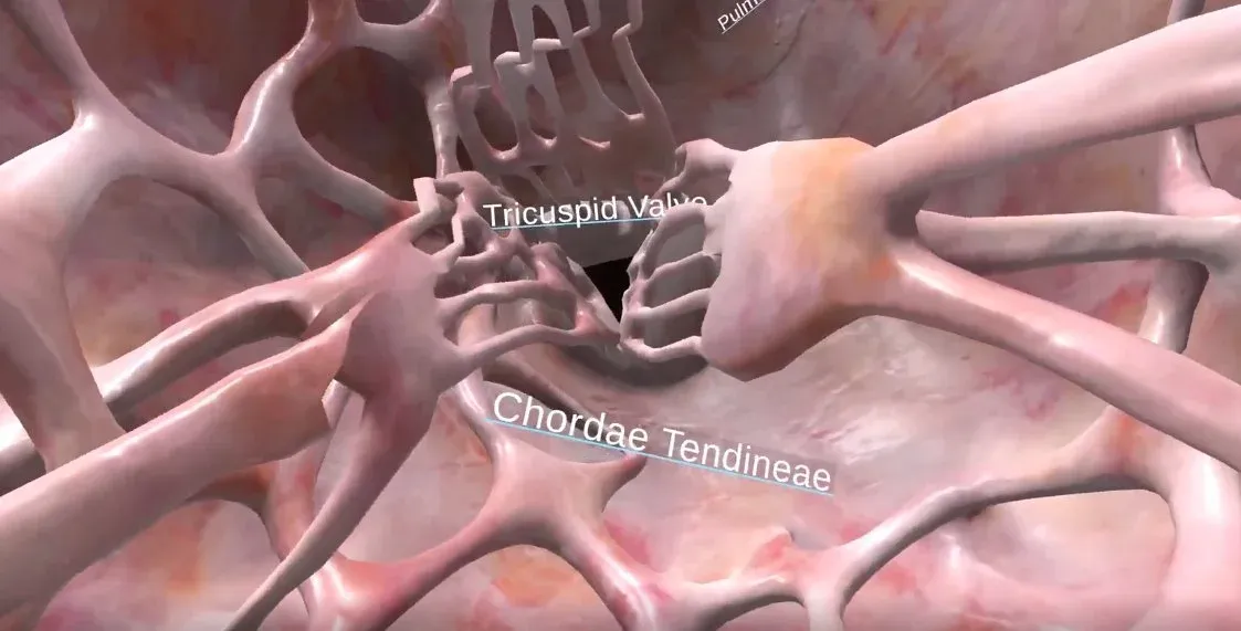
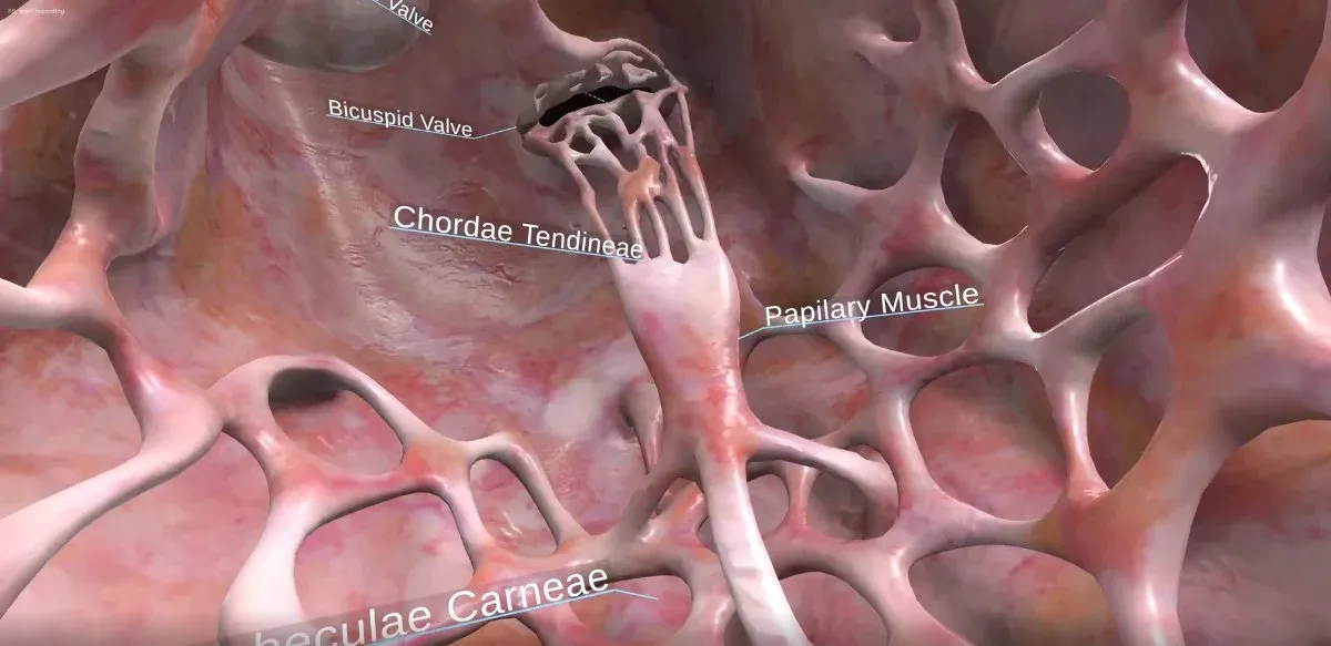
- Structures like chordae tendineae and papillary muscles only make sense once you can see them from inside the chamber.
- Moving between external, sectional and isolated views of the same organ builds a single coherent mental model.
- Blood flow is a path through space, so following it in space is the most direct way to learn it.
Structures you can identify
- Atria and ventricles
- Tricuspid (right) and bicuspid / mitral (left) valves
- Semilunar valves - aortic and pulmonary
- The interventricular septum
- Aorta, vena cava, pulmonary artery, and pulmonary veins
- Chordae tendineae, papillary muscles, and trabeculae carneae
Concepts it makes tangible
- The path of oxygenated and deoxygenated blood
- How the valves maintain one-way flow
- The link between chamber structure and function
- External, sectional, and isolated views of the same organ
- How anatomy underlies the cardiac cycle and circulation
How students explore it
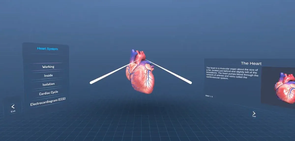
- A chapter hub moves between Working, Inside, Isolation, Cardiac Cycle, and ECG
- External anatomy is fully labelled
- Inside views open each chamber for close study
How faculty use it
- Pre-lab or lecture support for cardiovascular anatomy
- Self-paced revision of a complex 3D structure
- Demonstration for large groups
- Assessment of structures and blood flow
The 7thi AI tutor sits alongside the whole experience, scaffolding the difficult parts and giving subject-aware answers in context. Built-in assessment lets faculty see, per student, who has grasped which concepts - without grading another paper.
How the module works
Students explore the heart as a manipulable 3D model rather than a diagram. They rotate the external anatomy to identify the chambers, the great vessels and the surface features, then open the heart in section to study the valves and the internal wall structure, and finally step inside a chamber to see the chordae tendineae, papillary muscles and trabeculae carneae from within. A chapter hub moves between working, inside, isolation, cardiac-cycle and ECG views of the same organ, so students build one coherent mental model instead of stitching together separate pictures. Following oxygenated and deoxygenated blood through the chambers shows how the valves keep flow moving one way.
What students take away
A student can identify the atria, ventricles, the four valves, the septum and the great vessels, and locate internal structures such as the chordae tendineae and papillary muscles. They can trace the path of blood through the heart and explain how chamber structure relates to the pressure each side must generate - a solid three-dimensional grasp of cardiac anatomy that a flat diagram struggles to build.
In the classroom
Anatomy and physiology courses use the model for teaching, demonstration and revision. It lets a lecturer walk a whole group through the same structure at once, gives students a self-paced way to learn cardiac anatomy before assessment, and pairs naturally with the Cardiac Cycle module so that structure and function are learned together. Nursing, paramedic and allied-health programmes use it to build the spatial understanding that clinical examination later depends on.
Interactive features and modes
A chapter hub moves between working, inside, isolation, cardiac-cycle and ECG views of the same organ. Students can rotate the external anatomy, open the heart in section, isolate individual structures and step inside a chamber to see the valve apparatus from within. Blood flow can be followed through the chambers and great vessels, so structure and function are learned together rather than as separate facts.
Why it matters
The heart is a four-chambered three-dimensional pump, and yet it is most often taught from two-dimensional diagrams that force students to reconstruct its geometry mentally every time. Structures like the chordae tendineae and papillary muscles only make sense once they are seen from inside the chamber, and blood flow is fundamentally a path through space. A model students can rotate, section and step inside builds the spatial understanding that underpins everything from physiology to clinical examination.
The bigger picture
Cardiac anatomy is the foundation on which physiology, pathology and clinical skills are all built, and getting the three-dimensional structure wrong at the start makes everything that follows harder. A flat diagram forces students to reconstruct the geometry of a four-chambered pump in their heads every time, and internal structures like the chordae tendineae only make sense when seen from inside the chamber they occupy. A model that can be rotated, sectioned and entered gives students a durable spatial mental model of the heart, which is exactly what a diagram cannot.
Devices, deployment and assessment
Deploying the module is straightforward. It runs on Meta Quest, ClassVR and Pico headsets, on WebXR-compatible browsers, and on the desktop without any headset at all, so a department can start with the hardware it already has and scale as it grows. Faculty can assign the whole module or specific scenes, set it as pre-lab preparation or post-lab revision, and track progress on a dashboard that records concepts mastered, time per scene and assessment scores, with export to the LMS via xAPI. It pairs directly with the Cardiac Cycle module, so a department can teach structure and function as one connected experience. The 7thi AI tutor answers student questions in context throughout, so learners are never left stuck and faculty are not fielding the same question forty times.
Who this module is for
- Medical, nursing and allied health undergraduates studying cardiovascular anatomy
- Biology and physiology students covering the circulatory system
- Paramedic and health science programmes
- Faculty running anatomy revision or demonstration for large groups
Syllabus alignment
Where this module fits.
Nursing & Allied Health syllabi
Maps to foundational anatomy and physiology units in Nursing, Physiotherapy, Allied Health, Biomedical Science, and Life Sciences programmes.
NMC / CBME (India)
Indicatively supports anatomy and physiology learning outcomes related to cardiovascular structure, circulation, and function.
University syllabi
Maps to institution-specific anatomy and physiology syllabi, practical records, assignments, and assessment rubrics. Request a custom mapping.
Keep exploring
Related modules.
See Human Heart Anatomy live in a demo.
Thirty minutes, the full module, your curriculum questions answered.
Which structures can students identify?
Atria and ventricles, tricuspid and bicuspid valves, aortic and pulmonary semilunar valves, the interventricular septum, aorta, vena cava, pulmonary artery and veins, chordae tendineae, papillary muscles and trabeculae carneae.
Can students see inside the chambers?
Yes. Inside views open each chamber for close study, so the valve apparatus and the internal wall structure can be examined from within rather than inferred from a section.
How is blood flow demonstrated?
Students follow oxygenated and deoxygenated blood through the chambers and great vessels, seeing how the valves maintain one-way flow and how chamber structure relates to the pressure each side generates.
How does this relate to the Cardiac Cycle module?
This module covers structure; the Cardiac Cycle module animates one full beat. A chapter hub moves between Working, Inside, Isolation, Cardiac Cycle and ECG views, so the two are designed to be used together.
How long is a typical session?
About 20 minutes for a full run including assessment. Students can pause and resume, and faculty can assign specific parts rather than the whole module.
Which headsets does this module run on?
HTC, Meta Quest, ClassVR, WebXR, plus any WebXR-compatible browser. It also runs on desktop without a headset, which covers faculty preview and remote cohorts. OpenXR and 6DOF headsets are supported.
Is the assessment graded automatically?
Yes. Scores reach the faculty dashboard immediately, pass thresholds are adjustable, and results export to your LMS via xAPI.
Is this mapped to our curriculum?
It is mapped to ABET, NBA and equivalent frameworks, and we map it to your own cardiovascular system syllabus before deployment. Request a custom mapping.