
VR Module · Biology · Sciences
Internal structure of dicot & monocot - VR module.
Prepare and stain a thin section in VR, then examine it under the microscope - distinguish dicot from monocot stems and roots by their tissue layers and vascular bundle arrangement.
See it
Inside the module.
Learning objectives
By the end of this module, students will be able to:
Prepare a thin hand section and stain it with safranin to highlight lignified plant tissues
Mount the stained section and examine it under the compound microscope
Identify the tissue layers - epidermis, cortex, endodermis, vascular bundles, and pith
Distinguish a dicot stem (vascular bundles in a ring, open) from a monocot stem (scattered, closed)
Compare dicot and monocot roots by their xylem and phloem arrangement
Relate internal structure to function and to standard plant-anatomy theory
About the module
See the plan of a stem and root, layer by layer.
Comparing the internal structure of dicot and monocot stems and roots is a foundational plant-anatomy practical. This module recreates it in VR: students prepare a thin section, stain it, mount it, and study the arrangement of tissues under the microscope - the same workflow as a traditional botany lab, repeatable as often as they like.
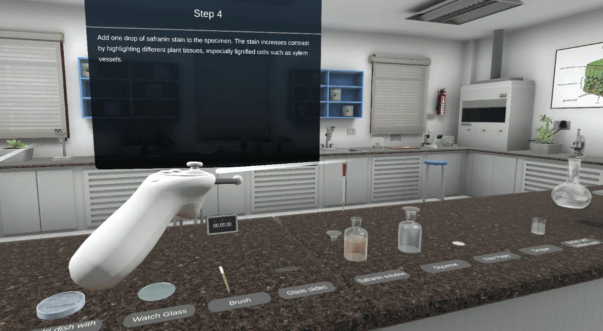
Learners are guided through each step - sectioning, adding a drop of safranin to raise contrast in lignified cells such as xylem vessels, mounting in glycerine, and viewing the slide. They then identify the epidermis, cortex, endodermis, vascular bundles, and pith, and read the tell-tale differences: a dicot stem shows vascular bundles in an open ring, while a monocot stem shows them scattered and closed.
Because it is virtual, students can repeat the preparation and comparison without consuming specimens, stains, or slides - ideal for pre-lab preparation, practical revision, and demonstrations. The 7thi AI tutor is on hand throughout, and built-in assessment shows faculty who has grasped each concept.
UGC / NEP 2020
Supports experiential, digital, multidisciplinary, and skill-based learning in Botany, Life Sciences, and Biology practicals - useful as a virtual lab component for plant anatomy.
NAAC / Outcome-Based Education
Evidences ICT-enabled teaching, repeatable practical work, and improved access to lab-based education, supporting practical learning outcomes and student engagement.
University syllabi
Maps to B.Sc. Botany, Life Sciences, and Plant Anatomy practical syllabi - including practical objectives, lab-record outcomes, viva preparation, and assessment rubrics. Request a custom mapping.
Lab Resources & Access
Reduces dependence on microscopes, stains, reagents, and prepared slides, giving every student unlimited, repeatable practice - useful where lab time or consumables are limited.
Get this module
Included in these iXRLabs packages.
Sciences
CoreCurriculum-aligned science VR modules across chemistry, physics, and biology.
Complete Sciences
All subjectsThe full sciences library, including Dicot & Monocot Structure.
Custom rollout
QuotePick specific modules, devices, and seat counts. We'll scope a deployment for your department.
7thi and assessment in this module
7thi in this module
Students can ask 7thi questions at any point - about dicot and monocot anatomy or anything they see in the section. 7thi answers in context, without breaking the flow.
Assessment
Session data - concepts mastered, time per scene, assessment scores - appears on the faculty dashboard. Export to your LMS via xAPI.
Keep exploring
Related sciences modules.

Do students need real slides, stains, or a microscope?
No. Sectioning, safranin staining, mounting, and microscope viewing are all simulated, so students can repeat the whole preparation with no consumables or equipment constraints.
What prior knowledge do students need?
Foundation-level biology. Suitable for B.Sc. Botany, Life Sciences, and Biology undergraduates from Year 1, and for pre-lab preparation before a wet lab.
How long is a typical session?
About 20 minutes for a full run including assessment. Students can pause and resume, and faculty can assign the stem or root comparison separately.
What does the module cover?
Section preparation and safranin staining, then identification of the epidermis, cortex, endodermis, vascular bundles, and pith - and the dicot-versus-monocot differences in both stem and root.
Which headsets is this module optimised for?
HTC, Meta Quest 2 / 3 / Pro, ClassVR, and any WebXR-compatible browser. It also runs in desktop browsers without a headset.
See the Dicot & Monocot module live in a demo.
Thirty minutes, the full module, your curriculum questions answered.